Biolog provides powerful cellular analysis tools for solving critical problems in pharmaceutical, industrial, agricultural and academic research and development. We are proudly present their key technologies here:
1) Microbial Identification
Biolog Microbial ID System can rapidly and accurately identify over 2,900 species of aerobic and anaerobic bacteria, yeasts and fungi. All Biolog Microbial Identification Systems — manual, semi-automated or fully automated — use the powerful new GEN III MicroPlate, allowing users to determine the most appropriate system to fit their current budget and level of throughput. Should needs change, all systems can be upgraded and expanded to meet new capacity requirements.
Fully automated high throughput aerobic identification
Semi-automated identification for bacteria, yeast, and filamentous fungi
Designed for teaching and low volume applications
Image source – https://www.biolog.com/products-portfolio-overview/microbial-identification/)
The new GEN III redox chemistry is applicable to an unprecedented range of both Gram-negative and Gram-positive bacteria. GEN III dissects and analyzes the ability of the cell to metabolize all major classes of biochemicals, in addition to determining other important physiological properties such as pH, salt, and lactic acid tolerance, reducing power, and chemical sensitivity.
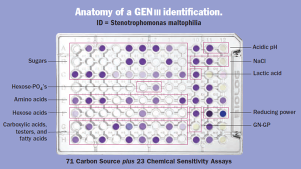
Image source – 00A 024 Rev D – Microbial Identification Systems Brochure)
2) Phenotype MicroArray for Microbial Cells & Mammalian Cells:
Phenotype MicroArray™ Technology enables researchers to characterize cells in up to 1,920 assays and evaluate cell changes under thousands of culture conditions and physiological states in a simple, rapid, efficient and cost-effective manner. By measuring a cell’s response to a genetic or environmental alteration, this integrated system of cellular assays, instrumentation, and bioinformatics software reveals invaluable information to speed insight and discovery and expedite scientific publication.
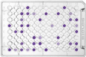
Phenotype MicroArrays are preconfigured sets of phenotypic tests deployed on microplate panels. Each well of the array is designed to test a different phenotype after inoculation with a standardized cell suspension, allowing simultaneous testing of thousands of phenotypes in a single experiment. PMs use Biolog’s patented redox technology, with cell respiration (NADH production) as a universal reporter. If the phenotype is strongly “positive” in a well, the cells respire actively, reducing a tetrazolium dye and forming a strong colour. If it is weakly positive or negative, respiration is slowed or stopped, and less colour or no colour is formed.
The redox assay provides for both amplification and precise quantitation of phenotypes. Incubation and recording of phenotypic data is performed automatically by the OmniLog® instrument, which captures a digital image of the MicroArray several times each hour and stores the quantitative colour change values into computer files. The computer files are then displayed in the form of kinetic graphs. Thousands of phenotypes are monitored simultaneously, and more than 470,000 data points can be generated in one 24 hour run. To compare the phenotypes of two cell lines, one is recorded as a red tracing and one as a green tracing. These graphs can then be overlaid by the bioinformatic software to detect differences. Areas of overlap (i.e. no change) are colored yellow, whereas differences are highlighted as patches of red or green.
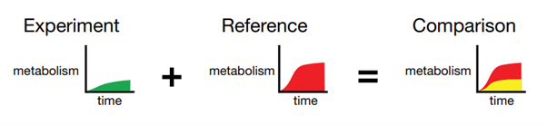
Image source – https://www.biolog.com/wp-content/uploads/2020/05/00A-037rA-PM-Microbiology-2011.pdf
Phenotype MicroArray technology enables researchers to evaluate nearly 2000 phenotypes of a microbial cell in a single experiment. Through comprehensive and precise quantitation of phenotypes, researchers are able to obtain an unbiased perspective of the effect on cells of genetic differences, environmental change, and exposure to drugs and chemicals. You can:
- Correlate genotypes with phenotypes
- Determine a cell’s metabolic and chemical sensitivity properties
- Discover new targets for antimicrobial compounds
- Optimize cell lines and culture conditions in bioprocess development
- Characterize cell phenotypes for taxonomic or epidemiological studies
For mammalian cells, Phenotype MicroArray technology can be used for diverse research and applications including:
- Correlating genotypes with phenotypes
- Studying metabolic reprogramming in cancer and anti-cancer drugs sensitivity
- Drug discovery research and toxicity screening
- Cell line and bioprocess improvement
- Cellular metabolism, metabolic disorders, nutrition
- Cell energies, growth and death
- Cell line QC and authentication
3) Microbiome:
Biolog has unique solutionsand choices for metabolic profiling of bacterial communities, as well as individual strains of interest, including those that are anaerobic or microaerophilic. See it in action below:
4) Ecoplate
Biolog EcoPlates provide a sensitive and reliable index of environmental change. This approach, called community-level physiological profiling (CLPP), is effective at distinguishing spatial and temporal changes in the metabolism of microbial communities. EcoPlates contain 3 repeated sets of 31 carbon sources and employ a tetrazolium redox dye as an indicator of microbial metabolism. As microbes utilize the carbon sources they respire and the tetrazolium reporter dye is reduced to form a visible purple color. Communities of microorganisms will exhibit a characteristic reaction pattern, a metabolic fingerprint, that reflects the metabolic properties of the community.

(Image source – https://www.biolog.com/wp-content/uploads/2020/05/00A-068-rA-EcoPlate-Brochure.pdf)
5) MitoPlate
Mitochondria play a primary role in energy production by cells. It is clear that these organelles are dynamic as the quantity and structure of the mitochondria in cells can change. Mitochondria are complex, consisting of over 1,000 proteins, the vast majority of which are coded for by nuclear rather than mitochondrial DNA. In addition to proteins, mitochondria also have specialized membranes and they can interact with each other and with other cellular organelles such as endoplasmic reticuli.
MitoPlates™ from Biolog provide a powerful research tool by allowing scientists to run preconfigured sets of 96 mitochondrial function assays in one experiment. Mitochondria can be interrogated and characterized in novel ways, looking at rates of substrate metabolism, sensitivity to drugs and other chemicals, and effects of mutations in mitochondrial genes.
Please watch the introductory video here:
6) Microbial Detection Using Chromogenic Media
Rainbow® Agars offer a simple, selective and chromogenic medium to help you conveniently detect strains of E. coli 0157, Salmonella, Shigella and Aeromonas with results in less than 24 hours.
RAINBOW AGAR O157
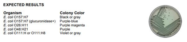
Rainbow Agar O157 has both selective and chromogenic properties that make it particularly useful for isolating pathogenic E. coli strains. The medium contains chromogenic substrates that are specific for two E. coli-associated enzymes: ßgalactosidase (a blue-black chromogenic substrate) and ß-glucuronidase (a red chromogenic substrate). Rainbow Agar O157 is listed in FDA’s Bacteriological Analytical Manual (BAM) and presents the agency’s preferred laboratory procedures for microbiological analyses of foods and cosmetics.
RAINBOW AGAR SALMONELLA
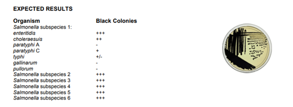
Rainbow Agar Salmonella utilizes an enhanced detection chemistry to determine H2S production among Salmonella spp.. Black colonies are formed by even weak H2S producing strains. In addition, novel selective agents increase the recovery rate of Salmonella while inhibiting the growth of other enteric bacteria and inhibiting H2S production by Citrobacter and other H2S positive enteric species.
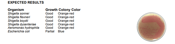
RAINBOW AGAR SHIGELLA / AEROMONAS
Rainbow Agar Shigella/Aeromonas was developed to provide laboratories with a better culture medium for directly isolating pathogenic strains of Shigella and Aeromonas. The medium is inhibitory to gram-positive bacteria and most non-enteric gram-negative bacteria, but is not toxic to the target species. Escherichia coli is significantly inhibited, and colonies that grow are blue.